Images scientifiques

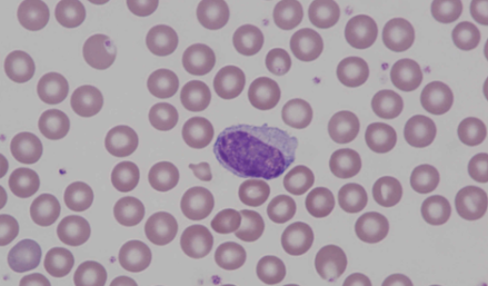
Typical lymphocyte of a mouse which is smooth and roundish with slightly rough nuclear chromatin, blue staining cytoplasm and a perinuclear halo.
<p>Typical lymphocyte of a mouse which is smooth and roundish with slightly rough nuclear chromatin, blue staining cytoplasm and a perinuclear halo.</p>

Two lymphocytes and an erythroblast (upper left). The erythroblast has a condensed, densely stained nucleus. The right lymphocyte is atypical in that the nucleus shows ruggedness and the nuclear chromatin pattern is slightly different compared to a normal lymphocyte. Two red blood cells with Howell-Jolly bodies (lower left).
<p>Two lymphocytes and an erythroblast (upper left). The erythroblast has a condensed, densely stained nucleus. The right lymphocyte is atypical in that the nucleus shows ruggedness and the nuclear chromatin pattern is slightly different compared to a normal lymphocyte. Two red blood cells with Howell-Jolly bodies (lower left).</p>

Monocytes show pleomorphic nuclei that may be round, indented or lobular. The extensive cytoplasm stains pale grey blue and frequently contains vacuoles.
<p>Monocytes show pleomorphic nuclei that may be round, indented or lobular. The extensive cytoplasm stains pale grey blue and frequently contains vacuoles. </p>

Murine (mouse) monocyte with notch
Many monocytes have a nucleus with a big notch. This cell shows another, smaller notch on the upper right of the nucleus. The cytoplasm shows the typical greyish colour.
<p>Murine (mouse) monocyte with notch </p> <p>Many monocytes have a nucleus with a big notch. This cell shows another, smaller notch on the upper right of the nucleus. The cytoplasm shows the typical greyish colour. </p>

Monocytes show pleomorphic nuclei that may be round, indented or lobular. The extensive cytoplasm stains pale grey blue and frequently contains vacuoles. This cell shows an atypical oval shape of the nucleus.
<p>Monocytes show pleomorphic nuclei that may be round, indented or lobular. The extensive cytoplasm stains pale grey blue and frequently contains vacuoles. This cell shows an atypical oval shape of the nucleus.</p>

Band neutrophil with ring-shaped nucleus. Band forms are infrequently seen in healthy rodents, but may be observed during inflammation.
<p>Band neutrophil with ring-shaped nucleus. Band forms are infrequently seen in healthy rodents, but may be observed during inflammation.</p>

Mouse neutrophils have pale cytoplasm with faint pink granules. The nucleus is typically highly segmented with threads connecting the nuclear segments.
<p>Mouse neutrophils have pale cytoplasm with faint pink granules. The nucleus is typically highly segmented with threads connecting the nuclear segments. </p>

