Images scientifiques

Polychromatic red blood cell (arrow). With immature red blood cells or deficient haemoglobin synthesis the red colour of haemoglobin and the blue staining of RNA become mixed to produce a polychromatic appearance. As the red blood cell matures and its haemoglobin content increases it appears more red.
<p>Polychromatic red blood cell (arrow). With immature red blood cells or deficient haemoglobin synthesis the red colour of haemoglobin and the blue staining of RNA become mixed to produce a polychromatic appearance. As the red blood cell matures and its haemoglobin content increases it appears more red.</p>

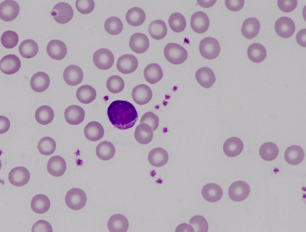
Small lymphocyte with slightly rough chromatin which stains dark purple. Purple azurophilic granules are present in the narrow band of cytoplasm.
<p>Small lymphocyte with slightly rough chromatin which stains dark purple. Purple azurophilic granules are present in the narrow band of cytoplasm. </p>

Blood smear of an aged rat (17-month-old) showing a picture of severe anaemia. In the centre, there is a neutrophil showing an atypically shaped nucleus, likely as a result of abnormal cell division.
<p>Blood smear of an aged rat (17-month-old) showing a picture of severe anaemia. In the centre, there is a neutrophil showing an atypically shaped nucleus, likely as a result of abnormal cell division.</p>


