Images scientifiques

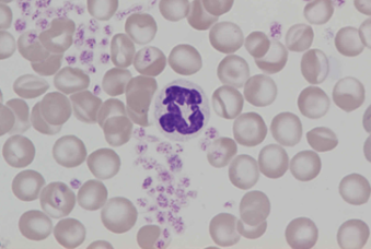
The nucleus of rat neutrophils is highly segmented, coiled, or ribbon-like with numerous indentations. The cytoplasm is pale with fine, diffuse granules. Mild platelet aggregation.
<p>The nucleus of rat neutrophils is highly segmented, coiled, or ribbon-like with numerous indentations. The cytoplasm is pale with fine, diffuse granules. Mild platelet aggregation.</p>

Rat neutrophils have pale cytoplasm with fine, diffuse granules. The nucleus is highly segmented, coiled, or ribbon-like with numerous indentations. This cell’s doughnut-shaped nucleus is folded.
<p>Rat neutrophils have pale cytoplasm with fine, diffuse granules. The nucleus is highly segmented, coiled, or ribbon-like with numerous indentations. This cell’s doughnut-shaped nucleus is folded.</p>

Anisocytosis, hypo- and polychromasia. In the centre, there is a neutrophil with a hypersegmented nucleus of which each segment is round – an atypical finding.
<p>Anisocytosis, hypo- and polychromasia. In the centre, there is a neutrophil with a hypersegmented nucleus of which each segment is round – an atypical finding.</p>

Blood smear of a rat showing various red blood cell morphologies: Howell-Jolly bodies (red arrow), helmet shapes, spherocytes, stomatocytes and a nucleated red blood cell.
<p>Blood smear of a rat showing various red blood cell morphologies: Howell-Jolly bodies (red arrow), helmet shapes, spherocytes, stomatocytes and a nucleated red blood cell.</p>

Blood smear of a rat showing marked anisocytosis, poikilocytosis and polychromasia. A nucleated red blood cell can be seen in the centre.
<p>Blood smear of a rat showing marked anisocytosis, poikilocytosis and polychromasia. A nucleated red blood cell can be seen in the centre.</p>

Typical monocyte of a rat. Both the colour shade of the cytoplasm and the shape of the nucleus reflect distinctive characteristics of this cell type.
<p>Typical monocyte of a rat. Both the colour shade of the cytoplasm and the shape of the nucleus reflect distinctive characteristics of this cell type.</p>

The first microscopically identifiable cell of granulocytic cell line.
Cell description:
Size: 12-20 µm
Nucleus: large, round or slightly oval with diffuse chromatin pattern and often 1-5 nucleoli
Cytoplasm: pale blue and usually agranular, sometimes Auer rods visible
<p>The first microscopically identifiable cell of granulocytic cell line. </p> <p>Cell description: </p> <p>Size: 12-20 µm </p> <p>Nucleus: large, round or slightly oval with diffuse chromatin pattern and often 1-5 nucleoli </p> <p>Cytoplasm: pale blue and usually agranular, sometimes Auer rods visible</p>

