Images scientifiques

Cell description:
Size: 15-25 µm
Nucleus: oval with identifiable nucleoli and diffuse chromatin structure Cytoplasm: basophilic with visible golgi-zone and eye-catching azurophil granula (primary granulation)
<p>Cell description: </p> <p>Size: 15-25 µm </p> <p>Nucleus: oval with identifiable nucleoli and diffuse chromatin structure Cytoplasm: basophilic with visible golgi-zone and eye-catching azurophil granula (primary granulation)</p>

Protubérances cytoplasmiques d'un lymphocyte dans le sang normal. (Bien que ces protubérances soient fréquemment présentes avec des infections, contrairement aux lymphocytes activés, elles ne sont d'aucune importance pour le diagnostic.)
<p>Protubérances cytoplasmiques d'un lymphocyte dans le sang normal. (Bien que ces protubérances soient fréquemment présentes avec des infections, contrairement aux lymphocytes activés, elles ne sont d'aucune importance pour le diagnostic.)</p>

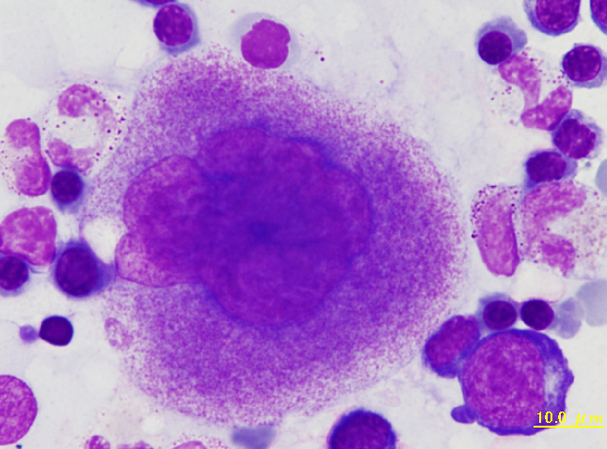
Bone marrow smear of a rabbit showing a megakaryocyte. The nucleus is lobulated and many azurophilic granules are found in the cytoplasm, suggesting maturity of the cell.
<p>Bone marrow smear of a rabbit showing a megakaryocyte. The nucleus is lobulated and many azurophilic granules are found in the cytoplasm, suggesting maturity of the cell. </p>

Eosinophil with large (approx. 1,5 µm) round and orange-reddish staining granules that are sharply defined and markedly larger than eosinophil granules in mice and rats.
<p>Eosinophil with large (approx. 1,5 µm) round and orange-reddish staining granules that are sharply defined and markedly larger than eosinophil granules in mice and rats.</p>

Two lymphocytes of a rabbit. The upper cell has a constricted nucleus which is more often seen with monocytes, but lacks the colour characteristic of monocyte cytoplasm.
<p>Two lymphocytes of a rabbit. The upper cell has a constricted nucleus which is more often seen with monocytes, but lacks the colour characteristic of monocyte cytoplasm.</p>


