Images scientifiques

Monocyte on top, heterophil below. Similar to eosinophils, the granules of leporine neutrophils stain intensely red by May-Giemsa or Wright staining. Therefore, they are termed pseudoeosinophils or heterophils in the rabbit.
<p>Monocyte on top, heterophil below. Similar to eosinophils, the granules of leporine neutrophils stain intensely red by May-Giemsa or Wright staining. Therefore, they are termed pseudoeosinophils or heterophils in the rabbit.</p>

Typical monocyte to the left and tri-segmented heterophil to the right. Similar to eosinophils, the granules of leporine neutrophils stain intensely red by May-Giemsa or Wright staining. Therefore, they are termed pseudoeosinophils or heterophils in the rabbit.
<p>Typical monocyte to the left and tri-segmented heterophil to the right. Similar to eosinophils, the granules of leporine neutrophils stain intensely red by May-Giemsa or Wright staining. Therefore, they are termed pseudoeosinophils or heterophils in the rabbit.</p>

Small lymphocyte with little cytoplasm to the left and a large atypical lymphocyte with abundant, intensely blue cytoplasm to the right.
<p>Small lymphocyte with little cytoplasm to the left and a large atypical lymphocyte with abundant, intensely blue cytoplasm to the right.</p>

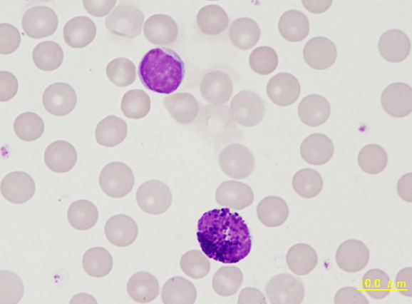
Small lymphocyte and a basophil of a rabbit. The basophil granules are well defined and numerous, almost concealing the outline of the nucleus similar to a mast cell.
<p>Small lymphocyte and a basophil of a rabbit. The basophil granules are well defined and numerous, almost concealing the outline of the nucleus similar to a mast cell. </p>

From left to right a band neutrophil, basophil and lymphocyte. In this basophil the basophilic granules are dense, but each granule is blurred, a common finding in rabbit basophils.
<p>From left to right a band neutrophil, basophil and lymphocyte. In this basophil the basophilic granules are dense, but each granule is blurred, a common finding in rabbit basophils.</p>

Giemsa simple stained blood smear of a rabbit showing a heterophil (neutrophil) to the upper left, a lymphocyte below and an eosinophil to the right. Note the lighter staining nucleus of the eosinophil as well as a lighter colour of the neutrophilic granules resulting from this stain.
<p>Giemsa simple stained blood smear of a rabbit showing a heterophil (neutrophil) to the upper left, a lymphocyte below and an eosinophil to the right. Note the lighter staining nucleus of the eosinophil as well as a lighter colour of the neutrophilic granules resulting from this stain.</p>

Trophozoites (rings) of Plasmodium falciparum are often thin and delicate, measuring on average 1/5 the diameter of the red blood cell. Rings may possess one or two chromatin dots. They may be found on the periphery of the RBC and multiple-infected RBC are not uncommon. Ring forms may become compact or pleomorphic, depending on the quality of the blood or if there is a delay in making smears.
<p>Trophozoites (rings) of Plasmodium falciparum are often thin and delicate, measuring on average 1/5 the diameter of the red blood cell. Rings may possess one or two chromatin dots. They may be found on the periphery of the RBC and multiple-infected RBC are not uncommon. Ring forms may become compact or pleomorphic, depending on the quality of the blood or if there is a delay in making smears.</p>

Sang périphérique (coloration de May-Grünwald-Giemsa) d'un patient de 27 ans atteint d'une leucémie myéloïde chronique (LMC). Globules rouges normaux, thrombocytose marquée (2750000 / µL), augmentation des granulocytes basophiles (->) et nombre normal de globules blancs sont observés. Dans ce cas, aucune mutation de JAK2 ne peut être détecté; à la place le patient a été testé positif pour le gène de fusion BCR-ABL.
<p>Sang périphérique (coloration de May-Grünwald-Giemsa) d'un patient de 27 ans atteint d'une leucémie myéloïde chronique (LMC). Globules rouges normaux, thrombocytose marquée (2750000 / µL), augmentation des granulocytes basophiles (->) et nombre normal de globules blancs sont observés. Dans ce cas, aucune mutation de JAK2 ne peut être détecté; à la place le patient a été testé positif pour le gène de fusion BCR-ABL.</p>